Observing Modes
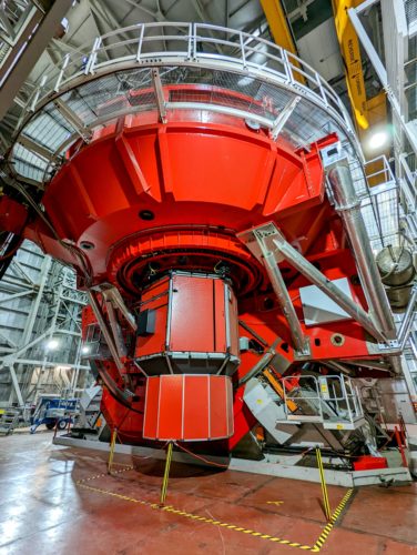
MODS1 as seen from chamber floor – S Allanson, LBTO
Direct Imaging
A set of SDSS filters is available in each MODS for Direct imaging. The following summarizes the imaging performance.
| Wavelength Range | 320-1100nm (Atmospheric to CCD limits) |
|---|---|
| Field of View | ~6.0×6.0 arcminutes (~2800×2800 pixels) |
| CCD Pixel Scale | Blue: 0.120 arcsec/pixel Red: 0.123 arcsec/pixel |
| Imaging Filters | SDSS u,g (Blue Channel) and SDSS r,i,z (Red Channel) |
| Image Quality | Best IQ within inner 2 arcmin, Distortions grow as r2 |
Long-slit Spectroscopy
Each MODS has a set of segmented long slits (5 60-arcsec long segments with a 3″ gap in between). The slit can be maintained at any position angle. The 0.6 arcsec wide slit will take advantage of the typical median seeing at the LBT and projects to ~5 pixels at the detector. Below summarizes the long-slit performance:
| Nominal Wavelength range | 320-1100nm |
|---|---|
| Red/Blue Dichroic Cross-Over | 565nm |
| Segmented Long-Slits | 0.3-arcsec Segmented long-slit 0.6-arcsec Segmented long-slit 0.8-arcsec Segmented long-slit 1.0-arcsec Segmented long-slit 1.2-arcsec Segmented long-slit 2.4-arcsec Segmented long-sli 60 x 5-arcsec long-slit |
| Diffraction Gratings | Red: 250 l/mm (R=2300) Blue: 400 l/mm (R=1850) |
| Double-Pass Prisms | Red: TIH6 + Ag (R=500-200) Blue: Fused Silica + Al (R=500-125) |
Multi-Slit Spectroscopy
Multi-object spectroscopy is accomplished with user-designed laser-machined slit masks loaded into a 24- position mask cassette. In multi–slit mode, a customized slit mask will be used to precisely locate a series of small and precisely positioned slits or apertures centered on targets within the MODS field of view. The full width of the grating spectrum is ~5000 pixels. Even with displaced slitlets the entire spectra will land on the detector (unlike with LUCI).
The performance is comparable to that of the long-slit, since the same detector, dichroic, gratings or prisms, filters, etc are used. The difference lies in that the image quality is dependent on the slit placement in the field. MODS is designed to deliver good images inside the central 4×4′ field of view and reduced image quality in a 6×6′ “extended” science field. In practical terms, the image quality decreases rapidly outside a 5.6-arcminute diameter circle, primarily due to a combination of astigmatism from the off-axis paraboloid collimator mirrors and off-axis aberrations in the LBT f/15 direct Gregorian focal plane. For purposes of locating MOS mask slits and alignment stars, it is recommended that primary science targets slits are kept within a 5.6′ circle, and alignment stars within a 5.0′ circle. Details on Mask design and optimization are described in the Mask Preparation Section.
Pre-defined (default) configurations:
There are four basic operating modes: acquisition, imaging, grating spectroscopy, and prism spectroscopy. Within in each mode, MODS may be configured for dual-channel (red+blue), blue-only, and red-only operation. The instrument configuration also sets the basic CCD subframe readout for that mode.
Each of the standard operating modes for MODS has a default instrument configuration. A summary of these default configurations or “Auto Configurations” is summarized below.
| Observing Configuration |
Mode | Active Channel | Grating | Filter | ROI |
|---|---|---|---|---|---|
| Long-Slit and Multi-Slit Spectroscopy |
Dual Grating | Red | G670L | Clear | 8K x 3K |
| Blue | G400L | Clear | 8K x 3K | ||
| Red Grating | Red | G670L | GG495 | 8K x 3K | |
| Blue Grating | |||||
| Blue | G400L | Clear | 8K x 3K | ||
| Dual Prism | Red | P700L | GG495 | 4K x 3K | |
| Blue | P450L | Clear | 4K x 3K | ||
| Red Prism | Red | P700L | GG495 | 4K x 3K | |
| Blue Prism | |||||
| Blue | P450L | Clear | 4K x 3K | ||
| Direct Imaging |
Dual Imaging | Red | flat | SDSSr, SDSSi, SDSSz |
3K x 3K |
| Blue | flat | SDSSg, SDSSu |
3K x 3K | ||
| Red Imaging | Red | flat | SDSSr, SDSSi, SDSSz |
3K x 3K | |
| Blue Imaging | |||||
| Blue | flat | SDSS g, SDSS u |
3K x 3K | ||
| Long-Slit and Multi-Slit Spectroscopy |
Red Acquisition | Red | flat | SDSSr, SDSSi, SDSSz |
1K x 1K |
| Blue Acquisition | |||||
| Blue | flat | SDSSg, SDSSu |
1K x 1K |
For example: When making and running the MODS Target Acquisition script, the default is the Red Camera, 60s r_sdss filter. Long Slit acquisition images are the central 1K x 1K ROI (Central 2 arcmin) and MOS acquisition images are the full 3K x 3K. When generating the acquisition script with the OT or with:
>> mkMODSAcq [options] rootName
options can be used to change the standard default configuration.
The options and how to use them when preparing scripts are outlined in the MODS Observing Script Preparation Toolkit page.